Medical Imaging
Medical imaging has different methods to create images of the human body in clinical purposes. Medical imaging is extremely important in confirming, correctly assessing and documenting the course of disease, and in assessing response to treatment.
Medical imaging services offered by our Center include :
- computed tomography (CT)
- magnetic resonance imaging (MRI)
- digital radiography
- digital mammography
- osteodensitometry
- ultrasound
- endoscopy
- elastography
Medical imaging (photo gallery)
- Doctors
- Medical services & prices

Radiology Senior Specialist
Medical Imaging Department Coordinating

Radiology Medical Imaging Specialist
Radiology Medical Imaging Senior Specialist
Radiology Medical Imaging Senior Specialist

Radiology Medical Imaging Senior Specialist
General Ultrasound Certified

Radiology Medical Imaging Specialist
Dr. Liliana Ştefan
Cardiology Specialist

Dr. Ramona Bică
Cardiology Senior Specialist

Dr. Angelica Nour-Dincă
Internal Medicine Senior Specialist
Cardiology Specialist
GP Specialist
Md/PhD
Dr. Lavinia Raluca Ene
Nephrology Specialist
Dr. Magdalena Zidu
Internal Medicine Senior Specialist, Rheumatology Specialist

Dr. George Sebastian Gherlan
Infectious Diseases Senior Specialist
Md/PhD
Lecturer

Dr. Mărioara Casian
Gastroenterology Senior Specialist
General abdominal ultrasound and digestive diagnostic endoscopy certified
Dr. Anna-Maria Tănase
General Surgery Senior Specialist

Dr. Florentina Mehic
Internal medicine Specialist
Dr. Aliona Chiriţă
Ginecology and Obstetrics Specialist

Dr. Cristina Daniela Ene
Endocrinology Senior Specialist
Dr. Radu-Ioan Vărşăndan
Urology & Andology Senior Specialist
Dr. Marinela Circa
Cardiology Senior Specialist
Doppler Ultrasound certified
Dr. Gabriela Scurtu
Pediatrics Senior Specialist
| RON | |
| Elastography | |
| Mamar elastography | 200 |
| Digital mammography | |
| Unilateral mammography | 200 |
| Bilateral mammography | 300 |
| Digital mammography - additional incidences | 60 |
| Imaging consultation (comparison with previous examinations, correlated analysis of data with other imaging methods, discussions and patient guidance, second opinion on mammograms performed in other medical institutions) | 200 |
| Digital radiology | |
| One radiographic incidence, any segment | 100 |
| Two radiographic incidences, large segments (skull, lungs, spine, pelvis, etc.) | 170 |
| Two radiographic incidences, small joints (knee, ankle, elbow, etc.) | 150 |
| To any additional over two incidences, 70 lei are added to each large segments and 50 lei each small joints. | |
| Ultrasound | |
| Echocardiography | 300 |
| Upper abdomen ultrasound | 200 |
| Lower abdomen ultrasound | 200 |
| General abdominal ultrasound | 300 |
| Testicle ultrasound | 150 |
| Thyroid ultrasound | 200 |
| General abdominal ultrasound - Acuson S2000 (about Acuson S2000) | 300 |
| Urinary tract ultrasound | 200 |
| Scrotal ultrasound | 150 |
| Pregnancy dating ultrasound examination (7 - 11 weeks) - transvaginal | 200 |
| Pregnancy dating ultrasound examination (7 - 11 weeks) - twin pregnancy - transvaginal | 300 |
| Transvaginal ultrasound examination | 200 |
| Ovulation monitoring ultrasound (2 - 3 transvaginal ultrasound) | 400 |
| First trimester (7 - 11 weeks) 4D ultrasound examination screening - morphology, biometry, Doppler | 350 |
| First trimester (7 - 11 weeks) 4D ultrasound examination screening - twin pregnancy - morphology, biometry, Doppler | 600 |
| 4D ultrasound examination 15 - 18 weeks (for Triple test) - morphology, biometry, Doppler | 400 |
| 4D ultrasound examination 15 - 18 weeks (for Triple test) - twin pregnancy - morphology, biometry, Doppler | 700 |
| Second / Third trimester (22 - 36 weeks) 4D ultrasound examination - morphology, biometry, Doppler | 600 |
| Second / Third trimester (22 - 36 weeks) 4D ultrasound examination - twin pregnancy - morphology, biometry, Doppler | 1 100 |
| Doppler ultrasound of lower limb arteries, bilateral | 300 |
| Doppler ultrasound of lower limb veins, bilateral | 300 |
| Doppler ultrasound of carotid arteries | 300 |
| Doppler ultrasound of lower limb arteries + veins, bilateral | 500 |
| Doppler ultrasound of lower limb arteries, unilateral | 200 |
| Doppler ultrasound of lower limb veins, unilateral | 200 |
| Doppler ultrasound of lower limb arteries + veins, unilateral | 300 |
| Doppler ultrasound of upper limb arteries + veins, bilateral | 500 |
| Doppler ultrasound of superior limb arteries, unilateral | 150 |
| Doppler ultrasound of upper limb veins, unilateral | 150 |
| Doppler ultrasound of upper limb arteries + veins, unilaterally | 250 |
| Breast ultrasound - bilateral - Acuson S2000 (about Acuson S2000) | 350 |
| Breast ultrasound - unilateral - Acuson S2000 (about Acuson S2000) | 250 |
| Breast ultrasound - unilateral (Acuson S2000) (women with mastectomy) | 300 |
| Breast ultrasound - silicone implant breast - bilateral | 400 |
| Breast ultrasound - silicone implant breast - unilateral | 250 |
| Breast ultrasound - children up to 12 years | 200 |
| Targeted mammary ultrasound (lesion / axilla) | 200 |
| Collection of nipple secretion | 50 |
| Breast ultrasound - extraprogram fee (requests on Saturdays / rest leave) | 100 |
| Renal artery ultrasound | 100 |
| Musculoskeletal & soft tissues Ultrasound | |
| Articular ultrasound - a region | 250 |
| Articular ultrasound - two regions | 320 |
| Ultrasound of soft parts (skin, subcutaneous tissue / muscles) - one region (arm / forearm / hand / thigh / leg etc.) | 250 |
| Ultrasound of soft parts (skin, subcutaneous tissue / muscles) - two regions (arm / forearm / hand / thigh / leg etc.) | 320 |
| Endoscopy | |
| Upper endoscopy | 300 |
| Colonoscopy | 400 |
| Proctosigmoidoscopy | 350 |
| Colposcopy | 250 |
| Osteodensitometry | |
| One test | 60 |
| Two tests | 100 |
| Three tests | 150 |
Use of X-rays for diagnostic purposes in our Center
Radiological safety norms in diagnostic and interventional radiology practices, approved by CNCAN Order no. 173 / 16.10.2003, Cap. IX "Medical Exposure", Subcap. 9.1 "Responsibilities", Art. 107, paragraph b): "no patient shall be given a diagnostic medical exposure unless the exposure is prescribed by a medical practitioner;".
X-rays diagnostic examinations are not carried out in our center without the recommendation of a specialist doctor.
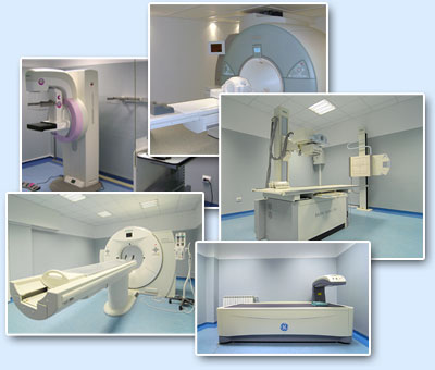
Medical imaging (photo gallery)
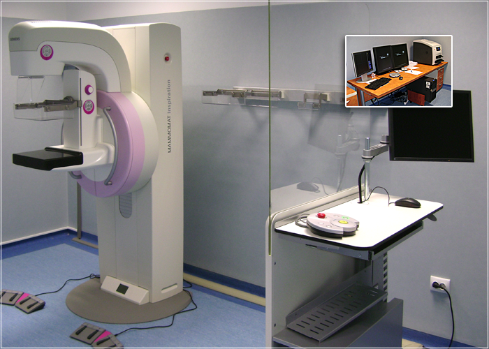
Mammography platform type MAMMOMAT Inspiration
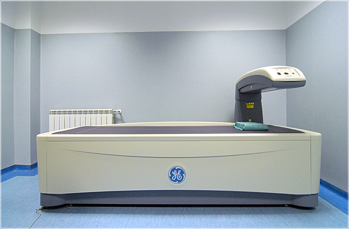
Bone Densitometer type General Electric - Prodigy Primo
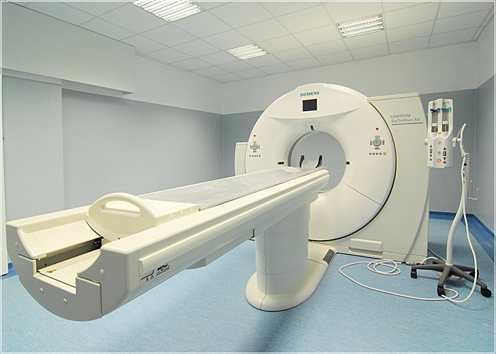
Computed tomography SOMATOM DEFINITION EDGE
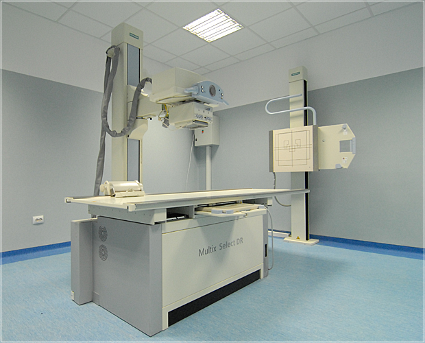
Digital X-ray Machine MULTIX SELECT DR
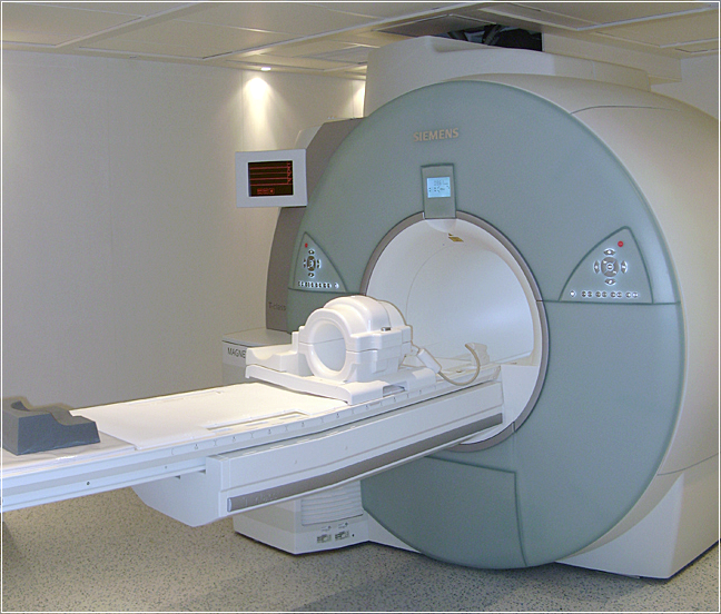
IRM equipment type MAGNETOM AVANTO FIT